![]() Figure 3 of
Johnson, Mol Vis 2007;
13:887-919.
Figure 3 of
Johnson, Mol Vis 2007;
13:887-919.
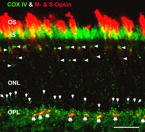
Figure 3. High magnification confocal image of cone juxtanuclear mitochondria and clusters of mitochondria in cone pedicles
Retinas were double labeled for COX IV (green) and the M- and S-cone opsins (red). The yellow-orange pixels show that COX IV and the cone opsins were in close apposition in the CIS and cone pedicles (double white arrows). Numerous large COX IV-positive juxtanuclear mitochondria are located above and below the distal cone nuclei (white arrowheads), while smaller COX IV-positive juxtanuclear mitochondria are located above rod nuclei (white arrows). Scale bar equal 20 μm.