![]() Figure 10 of
Johnson, Mol Vis 2007;
13:887-919.
Figure 10 of
Johnson, Mol Vis 2007;
13:887-919.
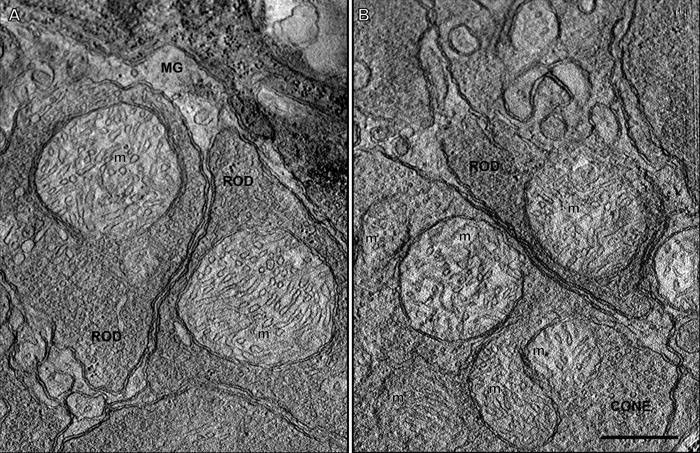
Figure 10. A 2.2 nm thick slice through a rod spherule and cone pedicle mitochondrion from three-dimensional electron tomographic reconstructions
The rod and cone mitochondria (m) are in the orthodox conformation, revealing that they were fixed and preserved in their non-energized state [55,123]. A: Two rod spherules present in the distal outer plexiform layer. Each rod spherule contains only one large almost circular mitochondrion. Müller glial processes (MG) encircle the rod spherules. B: A rod spherule overlying a cone pedicle in the outer plexiform layer. The larger cone pedicle contains five mitochondria (m). Scale bar equal 500 nm.