![]() Figure 5 of
Zhao, Mol Vis 2007;
13:873-880.
Figure 5 of
Zhao, Mol Vis 2007;
13:873-880.
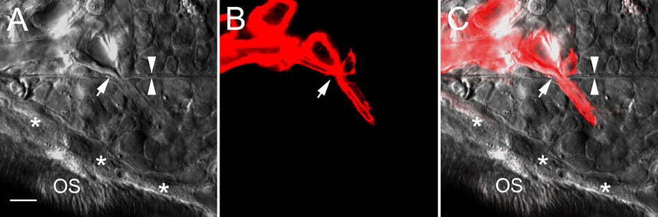
Figure 5. Choroidal neovascularization induced by Matrigel deposit
A: A differential interference contrast (DIC) image shows the posterior edge of a Matrigel deposit. A new blood vessel from the choriocapillaris had penetrated Bruch's membrane (between white arrowheads) at the site indicated by a white arrow. A new retinal pigment epithelium (RPE) layer (asterisks) had formed between the Matrigel deposit and photoreceptor outer segments (OS). B: The new blood vessel and the choriocapillaris are clearly shown in a confocal image (red). C: The DIC image in A superimposed on the confocal image in B. The scale bar represents 20 μm.