![]() Figure 3 of
Zhao, Mol Vis 2007;
13:873-880.
Figure 3 of
Zhao, Mol Vis 2007;
13:873-880.
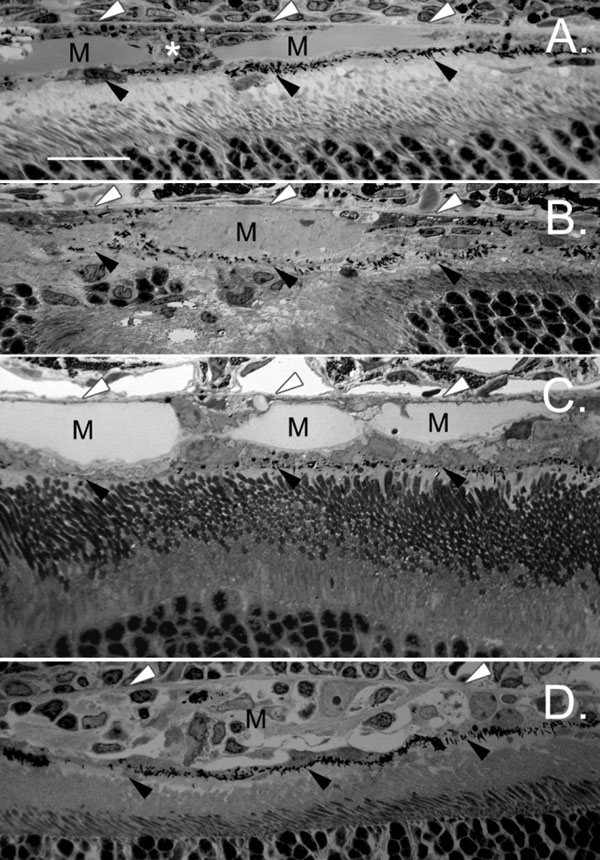
Figure 3. Formation of a new retinal pigment epithelium layer
A: A new monolayer (black arrowheads) formed between the Matrigel (M) and photoreceptors 10 days after Matrigel injection, displacing the Matrigel to the sub-RPE location. Comparable new retinal pigment epithelium (RPE) layers (black arrowheads) were seen (Bruch's membrane is indicated by white arrowheads) 25 days (B), 30 days (C), and 45 days (D) after Matrigel injection. In some cases, the Matrigel injected area (M) was filled with cells to form scare-like tissue (D). The scale bar represents 20 μm.