![]() Figure 5 of
Lopez-Martinez, Mol Vis 2007;
13:862-872.
Figure 5 of
Lopez-Martinez, Mol Vis 2007;
13:862-872.
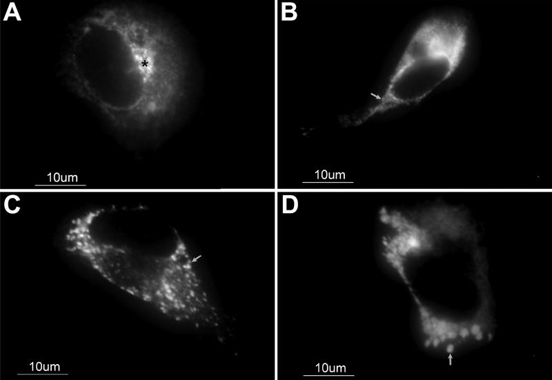
Figure 5. Subcellular distribution in transiently transfected 293T cells of human wild-type myocilin-GFP and two novel myocilin mutations found in this study
Two hundred nanograms of DNA constructs encoding wild-type myocilin (A), mutant myocilin forms, Arg346Thr (B) and Tyr479His (C), and the control, Pro370Leu (a disease causing mutation; D) were transfected into 293T cells. Wild-type myocilin was mainly detected in structures compatible with the Golgi apparatus and secretory vesicles. Note that the three mutant versions accumulated in the ER. The asterisk indicates the location of the Golgi apparatus. Arrows indicate the position of intracellular myocilin aggregates. Original magnification: X1600.