![]() Figure 1 of
Lopez-Martinez, Mol Vis 2007;
13:862-872.
Figure 1 of
Lopez-Martinez, Mol Vis 2007;
13:862-872.
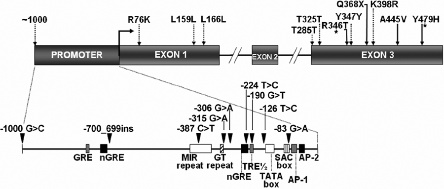
Figure 1. Genomic structure of the human myocilin gene and location of identified DNA sequence variations
The promoter and the three exons are represented by boxes. Consensus regulatory sequences in the promoter region are depicted in the inset. Pathogenic mutations and polymorphisms in the coding region are indicated by solid and dashed arrows, respectively. Sequence variations in the promoter region are shown by arrowheads. Novel mutations are shown by asterisks. All mutations are defined in terms of the one-letter code. Designated features include AP-1-like and AP-2-like sequences, putative TATA and SAC boxes, glucocorticoid response element (GRE), negative glucocorticoid response element (nGRE), thyroid response element (TRE) and a MIR repeat [26,63]. -700_699ins: -700_699insCAGACACACATATACATGCACATACACA.
