![]() Figure 5 of
Liu, Mol Vis 2007;
13:854-861.
Figure 5 of
Liu, Mol Vis 2007;
13:854-861.
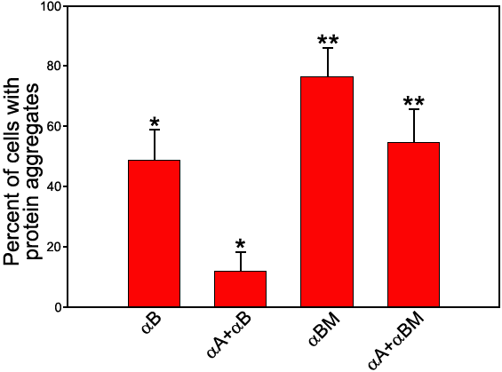
Figure 5. The percentage of cells with protein aggregates cotransfected with αA- and αB-crystallin or R120G αB-crystallin (αBM) genes
The x-axis labels are either RED-αB- or the pairs of GFP-αA- and RED-αB-crystallin. A four-fold decrease of cells with protein aggregates was observed for cotransfection of αA- and αB-crystallin (the asterisk indicates a p=0.006), but only about a 1.5 fold decrease for cotransfection of αA- and αBM-crystallin (the double asterisk indicates a p=0.02). The mean and standard deviation are shown as percentage ±SD and represent an average of three independent experiments. For each experiment, cells were counted in 30-50 confocal LSM images.