![]() Figure 9 of
Saito, Mol Vis 2007;
13:840-853.
Figure 9 of
Saito, Mol Vis 2007;
13:840-853.
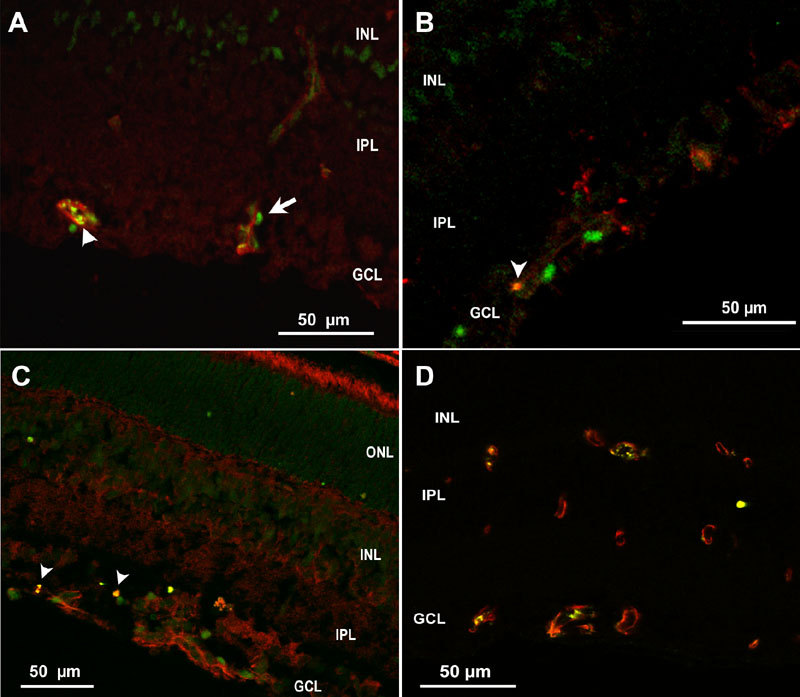
Figure 9. Cryosection of p18 oxygen-induced retinopathy eye from pups after intraperitoneal injection with phosphate buffered saline from p12 to p17
A: Cleaved caspase-3+ cells (green) were adjacent to lectin stained vessels (red), suggesting possible pericyte or leukocyte apoptosis, whereas lectin also co-stained with cleaved caspase-3 (arrowhead), supporting apoptosis of endothelial cells. B: Rare CD68+ stained cells (red) co-labeled with cleaved caspase-3 (Green) (arrowhead). C: Cleaved caspase-3+ nuclei in inner nuclear layer (INL) and ganglion cell layer (GCL, green), some of which only rarely co-labeled with anti-glial fibrillary acidic protein (GFAP, red arrowhead), suggesting apoptosis of glia. D: Mostly CD31+ endothelial cells (red) co-localized with gp91phox (green) in vessels. ONL represents outer nuclear layer; IPL represents inner plexiform layer.