![]() Figure 12 of
Saito, Mol Vis 2007;
13:840-853.
Figure 12 of
Saito, Mol Vis 2007;
13:840-853.
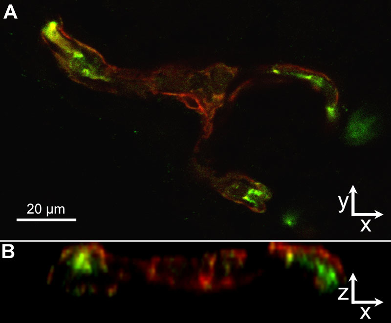
Figure 12. Cryosection of p18 oxygen-induced retinopathy eye from pups after intraperitoneal injections with phosphate buffered saline from p12 to p17
gp91phox localization (A, green) resided mainly inside of the /lectin stained (red) /vessel wall (z-series, B), suggesting that endothelial cells, and not NG-2 positive pericytes (red), express NAD(P)H oxidase in the oxygen-induced retinopathy model.