![]() Figure 10 of
Saito, Mol Vis 2007;
13:840-853.
Figure 10 of
Saito, Mol Vis 2007;
13:840-853.
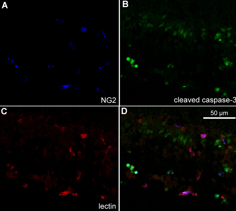
Figure 10. Cryosection of p18 oxygen-induced retinopathy eye from pups after intraperitoneal injection with phosphate buffered saline from p12 to p17
Labeling with NG2 (A, blue), cleaved caspase-3 (B, green), and lectin (C, red). Some NG2 labeled cells co-localized with cleaved caspase-3 (aqua, D) suggesting pericyte apoptosis. GCL represents ganglion cell layer; INL represents inner nuclear layer; IPL represents inner plexiform layer.