![]() Figure 2 of
Yu, Mol Vis 2007;
13:833-839.
Figure 2 of
Yu, Mol Vis 2007;
13:833-839.
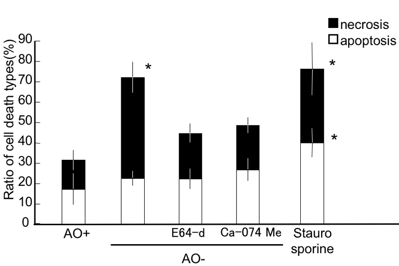
Figure 2. Detection of apoptotic and necrotic retinal ganglion cells under oxidative stress
Apoptotic retinal ganglion cells (RGCs) were significantly increased in the staurosporine-treated RGCs, but not in any other condition. Necrotic RGCs were significantly increased in the AO- and staurosporine conditions. E64-d and Ca-074 Me significantly reduced the necrotic cell percentage compared to the AO- conditions, but the values were not significantly different from the AO+ group. Each value represents mean±SD, (n=8). Asterisk indicates p<0.05, versus all the other groups (Tukey test).