![]() Figure 1 of
Yu, Mol Vis 2007;
13:833-839.
Figure 1 of
Yu, Mol Vis 2007;
13:833-839.
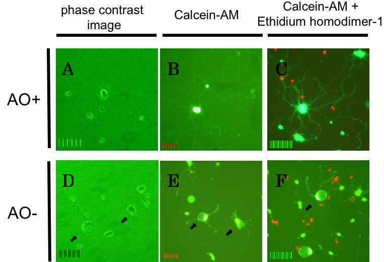
Figure 1. Rat retinal ganglion cell death by oxidative stress
Rat retinal ganglion cells (RGCs) after 72+24 h of culture under AO+ conditions (A, B, C) or AO- conditions (D, E, F). A, D: Phase contrast images of RGCs without labeling. B, E: Live cells with labeling of calcein-AM. C, F: Dead cells (red) with labeling of both calcein-AM and ethidium homodimer-1. The dendrites become shortened and cellular bodies had a deformed appearance (arrows). The scale bar represents 50 μm.