![]() Figure 4 of
Seigel, Mol Vis 2007;
13:823-832.
Figure 4 of
Seigel, Mol Vis 2007;
13:823-832.
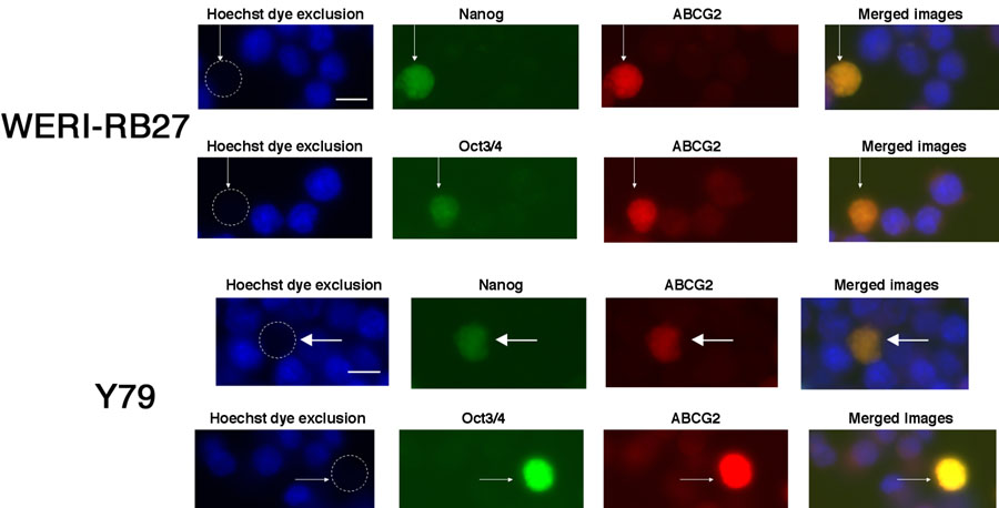
Figure 4. Colocalization of Nanog or Oct 3/4 in Hoechst-dim/ABCG2 positive cells
Y79 and WERI-RB27 human retinoblastoma cells were examined for fluorescent Hoechst 33342 dye uptake, ABCG2 immunoreactivity, coupled with either Nanog or Oct3/4 immunoreactivity. Each horizontal panel depicts the same microscopic field, viewed under separate fluorescent filters for Hoechst, FITC and TRITC, as well as a merged image of all three fields. As seen in the "Hoechst dye exclusion" field, the arrow points to a cell that has excluded the Hoechst dye and appears "Hoechst dim". This is due to the active Hoechst dye exclusion properties of the ABCG2 protein. In the next two panels, we see the same cell, as indicated by the arrow, that is immunoreactive for Nanog or Oct3/4 and ABCG2. When the three images are merged, ABCG2 colocalizes with both Nanog and Oct 3/4. The scale bar represents 5 μm.