![]() Figure 4 of
Li, Mol Vis 2007;
13:813-822.
Figure 4 of
Li, Mol Vis 2007;
13:813-822.
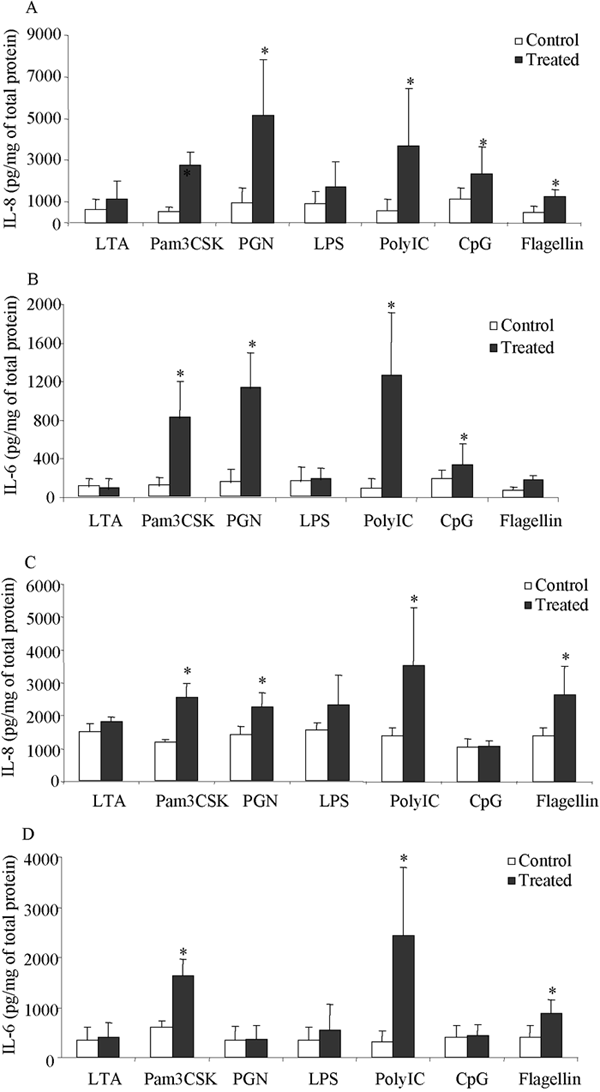
Figure 4. Ligand stimulated IL-6 and IL-8 secretion in cultured limbal and conjunctival epithelial cells
Eighty percent confluent conjunctival (A and B) and limbal (C and D) cells positive of corresponding TLR gene expression were incubated with different ligands at the concentrations indicated in Methods. Culture medium was collected 24 h later. IL-8 (A and C) and IL-6 (B and D) protein levels were measured by ELISA. Data represent the mean of four to five independent experiments and the error bar represents the SEM. The concentration of IL-6 and IL-8 was corrected by total protein amount in each well at the time of harvesting the supernatant. Significance compared to the controls by ANOVA and Fisher LSD analysis at a level of p<0.05 is shown by the asterisk. For CpG stimulated cells, the control refers to the CpG control DNA stimulated cells.