![]() Figure 3 of
Li, Mol Vis 2007;
13:813-822.
Figure 3 of
Li, Mol Vis 2007;
13:813-822.
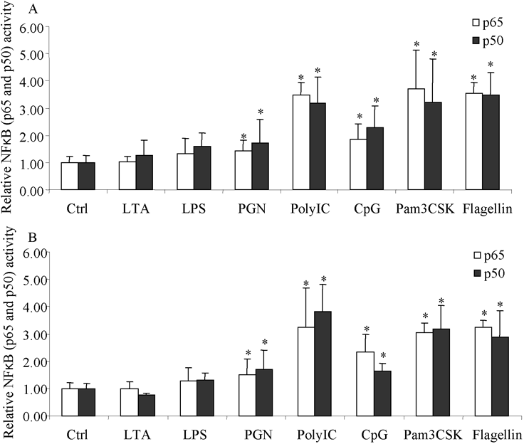
Figure 3. p50 and p65 activities in cultured limbal and conjunctival epithelial cells
Ligand stimulated p50 and p65 activities in cultured limbal (A) and conjunctival epithelial cells (B) are shown. Eighty percent confluent cells cultured in six well plates (positive for corresponding TLR gene expression) were incubated with different ligands at concentrations indicated in Methods for 8 h before harvesting the cells for the extraction of nuclear proteins. Equal amounts of nuclear proteins were used for the ELISA based analysis of p50 and p65 activities. p50 and p65 activities in control cells (without ligand stimulation) were set as 1 and used to normalize the activities measured in stimulated cells. The open bar represents the p65 subunit and the solid bar represents the p50 subunit. The results show the mean of three independent experiments and the error bar represents the SEM. The asterisk indicates a p<0.05 by ANOVA analysis followed with Fisher LSD test.