![]() Figure 4 of
Li, Mol Vis 2007;
13:804-812.
Figure 4 of
Li, Mol Vis 2007;
13:804-812.
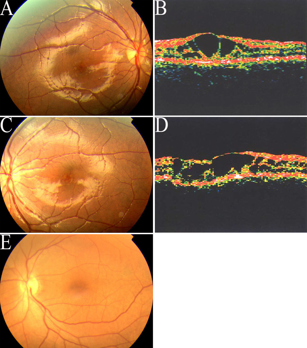
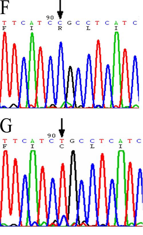
Figure 4. Fundus photographs, optical coherence tomography images, and DNA sequencing in Proband 140, and his grandfather, (Patient 142) of Family 140
The fundi of right (A) and left (B) eyes of Patient 140 showing cystoid-like maculopathies and golden-yellow reflex. Optical coherence tomography images of his right (C) and left (D) eyes revealed splitting had occured in the inner retina around the fovea. A normal fundus appearance (E) can be seen in his grandfather (Patient 142) with the same mutation. When ompared with the normal control (F), DNA sequencing showed the Arg200Cys mutation (598C->T) of exon 6 in Proband 140 (G).