![]() Figure 3 of
Li, Mol Vis 2007;
13:804-812.
Figure 3 of
Li, Mol Vis 2007;
13:804-812.
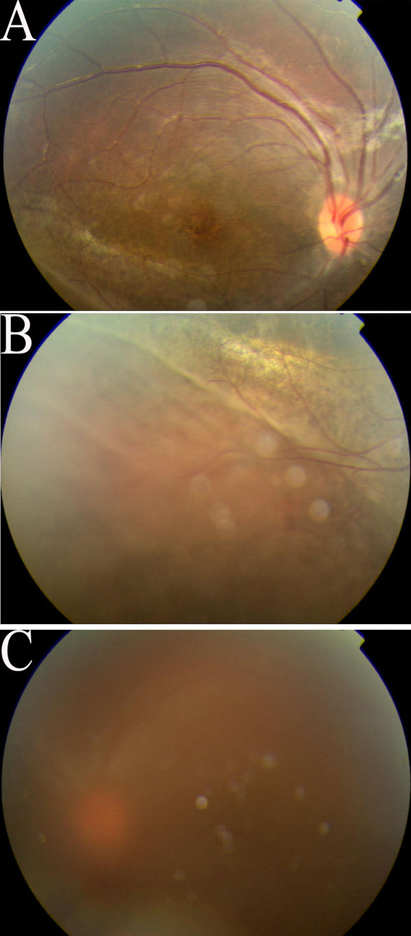
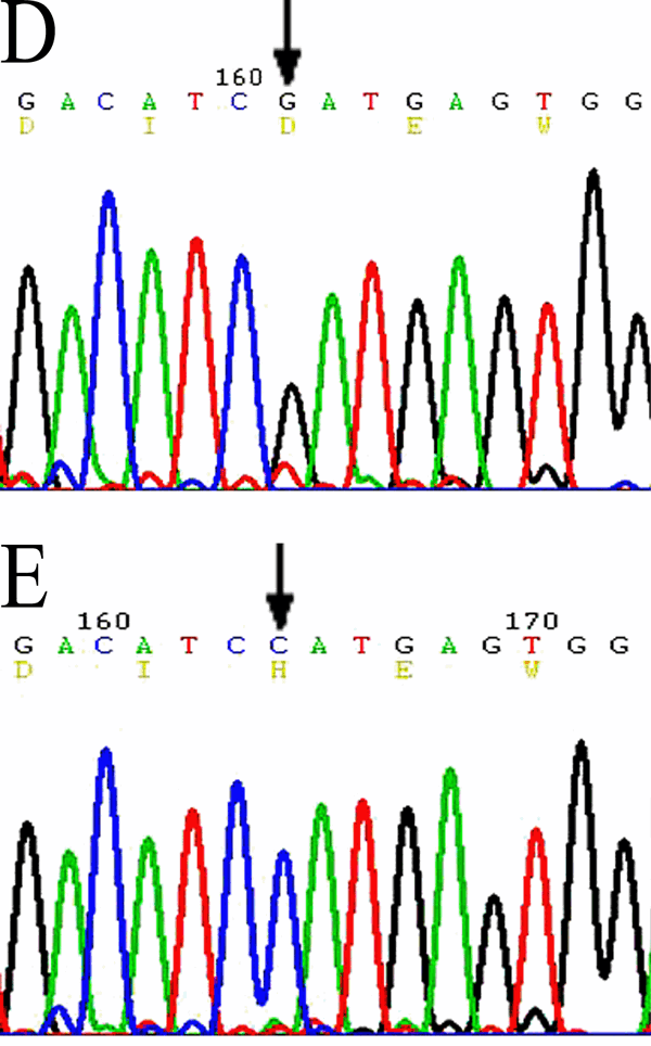
Figure 3. Fundus photographs and DNA sequencing in Patient 60 of Family 60
Fundus of right eye (A, B) showing cystoid-like maculopathy and peripheral retinoschisis. The left eye underwent vitrectomy with comobined sclera buckling because of retinal detachment and vitreous hemorrhage (C). Peripheral retinoschisis was found during the operation. DNA sequencing showed missense mutation -433G->C, Asp 145 His of exon 5 in normal control (D) and Patient 60 (E).