![]() Figure 2 of
Li, Mol Vis 2007;
13:804-812.
Figure 2 of
Li, Mol Vis 2007;
13:804-812.
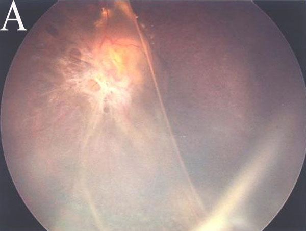
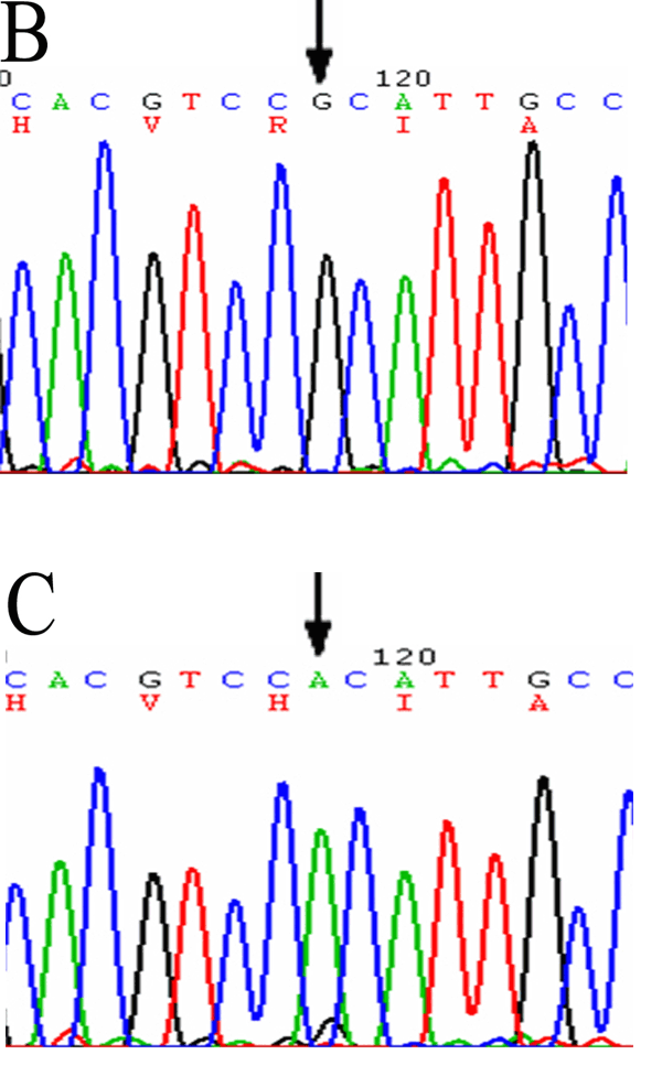
Figure 2. Fundus photographs and DNA sequence in Patient 20 of Family 20
Fundus of left eye (A) showing severe foveal and peripheral retinoschisis with tractional retinal detachment and proliferative vitreoretinopathy. DNA sequencing examination revealed Arg209His mutation -626G->A of exon 6 in normal control (B) and Patient 20 (C).