![]() Figure 4 of
Harris, Mol Vis 2007;
13:785-796.
Figure 4 of
Harris, Mol Vis 2007;
13:785-796.
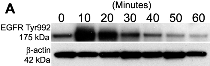
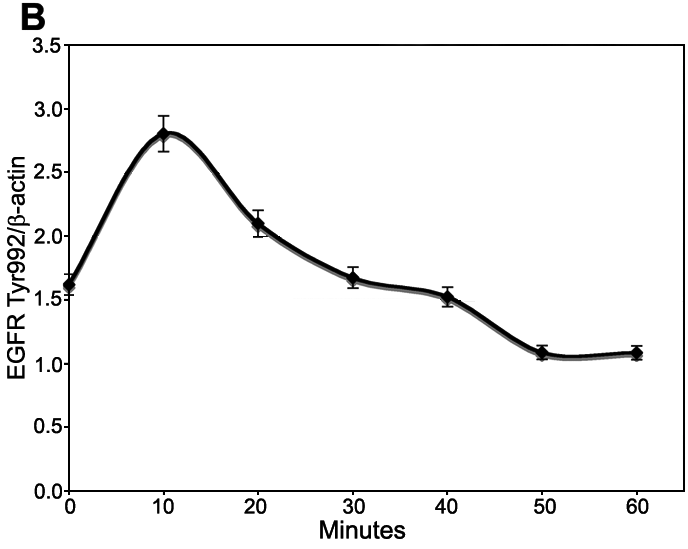
Figure 4. Time-course of EGFR Tyr992 phosphorylation
Subconfluent rat corneal endothelial cells were serum-starved and then treated with 25 ng/ml EGF in Medium 199 plus gentamicin. Samples were taken at various times for western blot analysis. A: Representative western blot showing the time-course of EGFR Tyr992 phosphorylation. Phosphorylated EGFR Tyr992 yields a 175 kDa band. β-Actin (42 kDa) was used for a loading control. B: Graph showing the average level of phosphorylated EGFR Tyr992 in duplicate gels from three separate experiments. β-Actin was used for normalization. Bars represent SEM.