![]() Figure 3 of
Harris, Mol Vis 2007;
13:785-796.
Figure 3 of
Harris, Mol Vis 2007;
13:785-796.
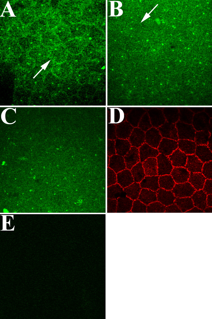
Figure 3. Fluorescent confocal images of EGFR localization in corneal endothelial cells of ex vivo rat corneas following EGF stimulation
In the absence of EGF, EGFR is located mainly at cell borders (arrow), although some cytoplasmic punctate staining is visible (A). After EGF stimulation for 15 min (B), EGFR staining is greatly reduced at cell borders (arrow). After 60 min (C), little-to-no EGFR is visible at cell borders; however, ZO-1 staining of the same tissue (D) indicates that cell borders remain intact. No staining is observed in the endothelium of ex vivo corneas incubated in secondary antibody alone (E). Note that small dots of intense stain can be observed scattered in the cytoplasm of cells in (A-C). The specific nature of this staining is unclear, since all antibodies were centrifuged at high speed prior to dilution to prevent nonspecific antibody deposition. Final magnification: 1000X.