![]() Figure 2 of
Harris, Mol Vis 2007;
13:785-796.
Figure 2 of
Harris, Mol Vis 2007;
13:785-796.
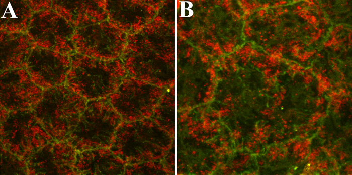
Figure 2. Representative fluorescent confocal images showing immunostaining for PTP1B and β-catenin in corneal endothelial cells of ex vivo rat corneas
PTP1B (red) localization is compared with that of β-catenin (green), a protein closely associated with cell-cell adhesion junctions. The staining pattern of PTP1B strongly suggests that PTP1B is associated with vesicular structures subjacent to the plasma membrane. The higher magnification image in (B) is included to more clearly demonstrate this localization pattern. The final magnification in A was 2100X and in B was 3120X.