![]() Figure 1 of
Suzuki, Mol Vis 2007;
13:772-778.
Figure 1 of
Suzuki, Mol Vis 2007;
13:772-778.
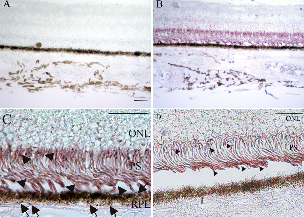
Figure 1. Immunohistochemical examination for oxidized phospholipids of a normal eye
A: Retinal section from the macula area of a normal eye. Negative control with non-immunized mouse IgM as a primary antibody. Original magnification, x100. Scale bar represents 50 μm. B: Retinal section from the macula area of a normal eye. Oxidized phospholipids are detected in the inner and outer retina and choroid. Original magnification, x100. Scale bar represents 50 μm. C: Retinal section from the macula area of a normal eye. Oxidized phospholipids are seen mostly in the photoreceptors (PS) and retinal pigment epithelium (RPE). Immunohistochemical analysis of the photoreceptor inner and outer segments demonstrated diffuse immunoreactivity (arrowheads). Both the basal and apical sides of the RPE cells are primarily stained (arrows). Original magnification, x600. Scale bar represents 50 μm. D: Retinal section from the peripheral area of a normal eye. Oxidized phospholipids are seen in the photoreceptor inner and outer segments (arrows). Immunoreactivity was less intense than in the macular area. Original magnification, x600. Scale bar represents 50 μm. ONL indicates outer nuclear layer