![]() Figure 3 of
Steele, Mol Vis 2007;
13:764-771.
Figure 3 of
Steele, Mol Vis 2007;
13:764-771.
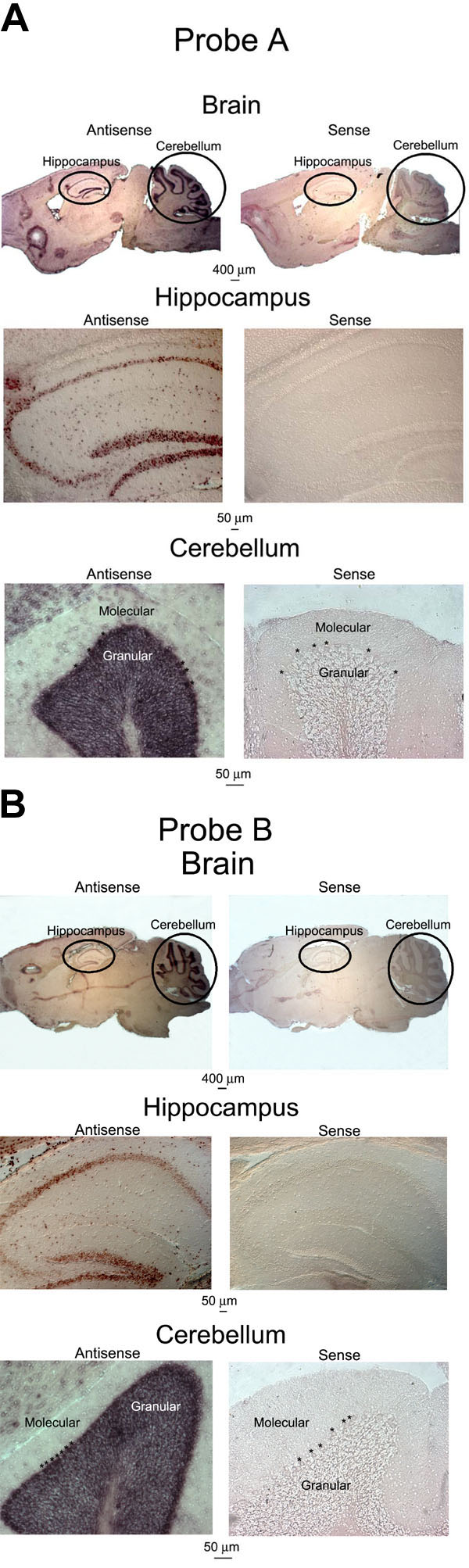
Figure 3. In situ hybridization analysis of mouse brain sections confirms specificity of Cav1.3 (α1D) cRNA probes
A: Brain sections from wild type C57BL/6J mice were hybridized with Probe A antisense and sense probes. Antisense probe (left panels) detected Cav1.3 (α1D) mRNA specifically in the hippocampus (middle panel) and cerebellum (bottom panel). In the cerebellum, strong labeling was observed in the large Purkinje neurons (large somas between molecular and granular cell layers, just below asterisks, *, in bottom panels) and in neurons within the granular layer, but was absent from cells of the molecular layer. Sense probes (right panels) did not detect Cav1.3 (α1D) mRNA in any cells of the hippocampus (middle panel) or cerebellum (bottom panel). The observation of such region-specific expression of Cav1.3 (α1D) mRNA in the brain supports the specificity of the Cav1.3 (α1D) probes. B: Brain sections from wild type C57BL/6J mice were hybridized with Probe B antisense and sense probes. Antisense probe (left panels) detected Cav1.3 (α1D) mRNA specifically in the hippocampus (middle panel) and cerebellum (bottom panel). In the cerebellum, strong labeling was observed in the large Purkinje neurons (large somas between molecular and granular cell layers, just below asterisks, *, in bottom panels) and in neurons within the granular layer, but was absent from cells of the molecular layer. Sense probes (right panels) did not detect Cav1.3 (α1D) mRNA in any cells of the hippocampus (middle panel) or cerebellum (bottom panel). The observation of such region-specific expression of Cav1.3 (α1D) mRNA in the brain supports the specificity of the Cav1.3 (α1D) probes.