![]() Figure 2 of
Steele, Mol Vis 2007;
13:764-771.
Figure 2 of
Steele, Mol Vis 2007;
13:764-771.
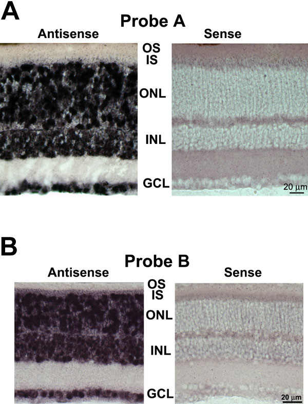
Figure 2. In situ hybridization analysis of Cav1.3 (α1D) mRNA expression in mouse retinal sections confirms expression in most retinal neurons, including rod photoreceptors
A: (Probe A) Retinal sections from wild type C57BL/6J mice were hybridized with A antisense and sense probes. Antisense probe (left panel) detected Cav1.3 (α1D) mRNA in most, if not all, cells of all three nuclear layers. Sense probe (right panel) did not detect Cav1.3 (α1D) mRNA in any cells of any nuclear layer. IS represents inner segments; OS represents outer segments; ONL represents outer nuclear layer; INL represents inner nuclear layer; GCL represents ganglion cell layer. B: (Probe B) In situ hybridization analysis of Cav1.3 (α1D) mRNA expression in mouse retinal sections confirms expression in most retinal neurons, including rod photoreceptors. Retinal sections from wild type C57BL/6J mice were hybridized with B antisense and sense probes. Antisense probe (left panel) detected Cav1.3 (α1D) mRNA in most, if not all, cells of all three nuclear layers. Sense probe (right panel) did not detect Cav1.3 (α1D) mRNA in any cells of any nuclear layer.