![]() Figure 1 of
Steele, Mol Vis 2007;
13:764-771.
Figure 1 of
Steele, Mol Vis 2007;
13:764-771.
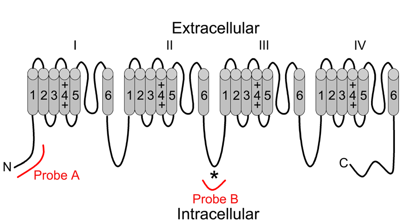
Figure 1. Topological correspondence of Cav1.3 (α1D) in situ hybridization probes
Voltage-dependent calcium channel pore-forming α1 subunits are composed of a single polypeptide consisting of 4 repeating domains (I-IV) joined by intracellular loops. Each repeat domain consists of 6 transmembrane-spanning regions (1-6) connected by intracellular and extracellular loops. Antisense and sense probe sets were generated using cRNA corresponding to 2 different topological regions of the Cav1.3 (α1D) subunit polypeptide. Probe A corresponded to cRNA encoding an N terminal fragment of the polypeptide, while B corresponded to cRNA encoding a fragment of the variable loop between repeat domains II and III. Probe B encompassed the antigenic peptide (*) used to generate the commercially available antibody used in most published immunohistochemical studies of Cav1.3 (α1D) expression.