![]() Figure 3 of
Liu, Mol Vis 2007;
13:758-763.
Figure 3 of
Liu, Mol Vis 2007;
13:758-763.
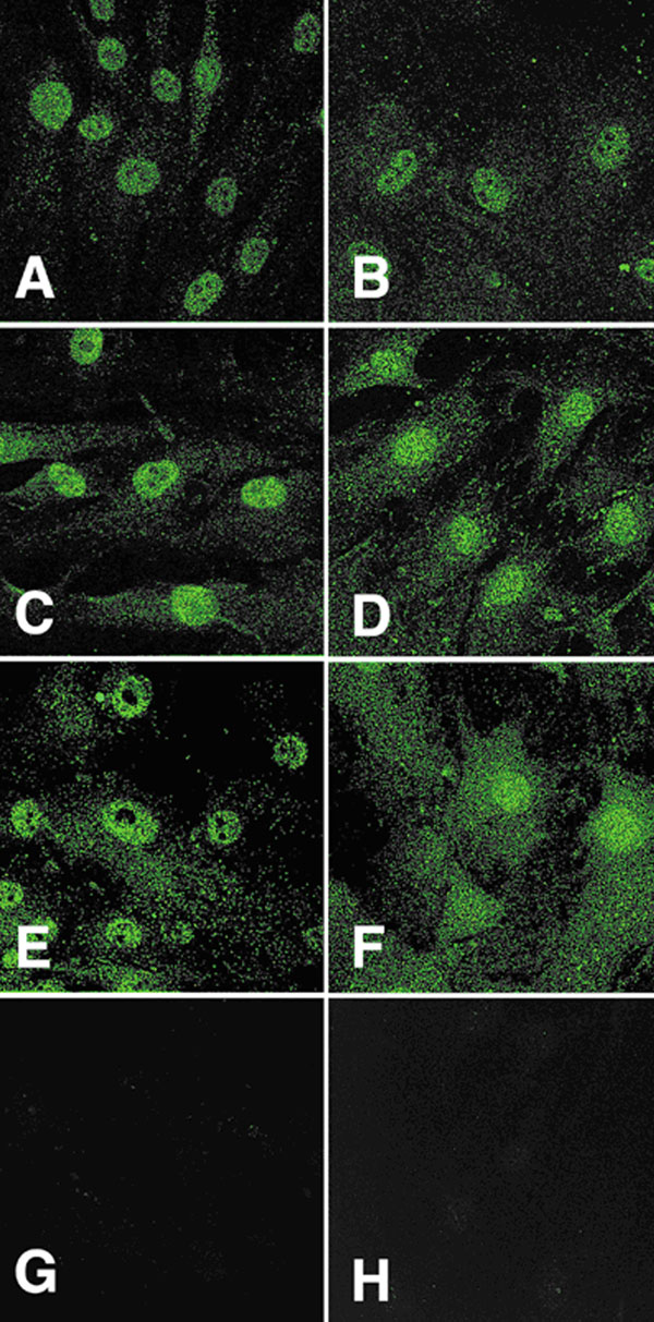
Figure 3. Immunofluorescent localization of ciliary neurotrophic factor tripartite receptor complex proteins in optic nerve head cells
Lamina cribrosa cells are in A, C, and E and optic nerve head astrocytes are in plates B, D, and F. A and B are examples of ciliary neurotrophic factor-α staining of the cells; plates C and D correspond to LIFR-β cellular staining; E and F are representatives of gp130 staining of both cell types; G and H are examples of negative controls (primary antibody omitted).