![]() Figure 6 of
Hartung, Mol Vis 2007;
13:66-78.
Figure 6 of
Hartung, Mol Vis 2007;
13:66-78.
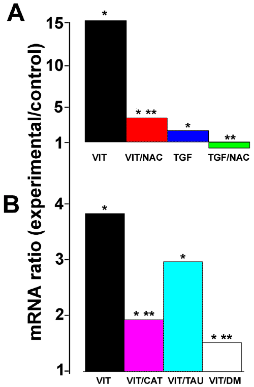
Figure 6. Reactive oxygen species quenchers inhibit the vitreous-mediated and transforming growth factor-β-mediated increase in heme oxygenase-1 mRNA expression
A: Retinal pigment epithelial (RPE) cells were incubated with control medium, vitreous-containing medium (VIT) or TGF-β1 (1 ng/ml)-containing medium for 12 h with or without 2 mM N-acetyl cysteine (NAC). HO-1 mRNA was measured by qPCR. All ratios are compared to the level in cells in control medium. B: RPE cells were incubated for 12 h in control medium, vitreous-containing medium (VIT) or vitreous-containing medium supplemented with 200 units catalase (VIT/CAT) per ml, 40 mM taurine (VIT/TAU) or 250 μM DMSO (VIT/DM). HO-1 mRNA was measured by qPCR. All ratios are relative to the level in cells in control medium (no change = ratio of 1.0). Asterisk (*) indicates that REST-XL analysis showed a change that is significantly different from control (p<0.05). Double asterisk (**) indicates a value significantly different from the level of HO-1 mRNA in vitreous-treated cells (p<0.05).