![]() Figure 5 of
Hartung, Mol Vis 2007;
13:66-78.
Figure 5 of
Hartung, Mol Vis 2007;
13:66-78.
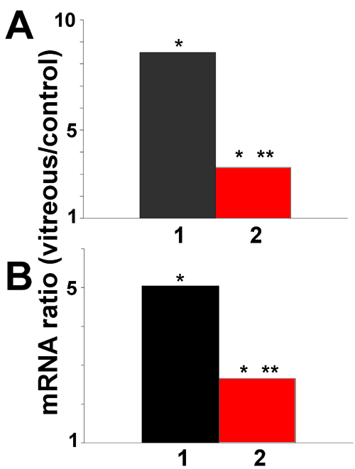
Figure 5. Inhibition of transforming growth factor-β signaling reduces the vitreous-mediated increase in heme oxygenase-1 expression by retinal pigment epithelial cells
A: Retinal pigment epithelial (RPE) cells were incubated for 12 h in vitreous-containing medium (bar 1) or in the same medium supplemented with SB431542 (10 μM), an inhibitor of type I TGF-β receptor signaling (bar 2). Heme oxygenase-1 (HO-1) mRNA expression was measured by qPCR. B: RPE cells were incubated in vitreous-containing medium (bar 1) or in the same medium supplemented with pan anti-transforming growth factor-β neutralizing antibody (10 μg/ml). HO-1 mRNA expression was measured by qPCR. Values show the change in mRNA expression compared to control (no change=1). Asterisk (*) indicates that REST-XL analysis showed a change that is significantly different from control levels of HO-1 mRNA (p<0.05). Double asterisk (**) denotes a value significantly different from the level of HO-1 mRNA in vitreous-treated cells (p<0.05).
