![]() Figure 4 of
Hartung, Mol Vis 2007;
13:66-78.
Figure 4 of
Hartung, Mol Vis 2007;
13:66-78.
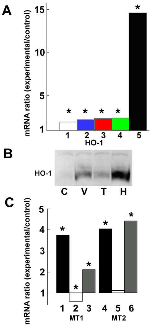
Figure 4. Effect of transforming growth factor-β1 on expression of heme oxygenase-1 and metallothioneins in retinal pigment epithelial cells
A: Retinal pigment epithelial (RPE) cells of the same donor and passage were incubated in control medium or with 25% vitreous or various concentrations (0.5 -10 ng/ml) of transforming growth factor-β1 (TGF-β1) for 12 h. Heme oxygenase-1 (HO-1) mRNA levels in vitreous- or TGF-β1-treated cells compared to control cells were measured by qPCR using ribosomal protein, large, P0 (RPLP0) mRNA levels to correct for any differences in the amount of cDNA. Bar 1: 0.5 ng TGF-β. Bar 2: 1 ng/ml. Bar 3: 5 ng/ml. Bar 4: 10 ng/ml. Bar 5: 25% vitreous. Values show the change in mRNA expression compared to control (no change =1). Asterisk (*) indicates that REST-XL analysis showed a change that is significantly different from control (p<0.05). B: RPE cells were incubated with normal medium (C), vitreous-containing medium for 24 h (V), TGF-β1 (5 ng/ml)-containing medium (T) for 24 h, or hemin-containing medium (H) for 24 h as a positive control. The proteins were analyzed by immunoblotting using rabbit anti-HO-1 antibodies. C: RPE cells were incubated with 25% vitreous (bars 1, 4) or 1 ng TGF-β1 per ml medium (bars 2, 5) or vitreous and TGF-β combined (bars 3, 6) for 12 h. mRNA for MT-1a (bars 1, 2, 3) and MT-2a (bars 4, 5, 6) in treated cells relative to untreated, control cells was determined. Asterisk (*) indicates that REST-XL analysis showed a change that is significantly different from control (p<0.05).