![]() Figure 3 of
Hartung, Mol Vis 2007;
13:66-78.
Figure 3 of
Hartung, Mol Vis 2007;
13:66-78.
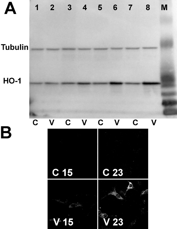
Figure 3. Expression of heme oxygenase-1 protein in vitreous-treated retinal pigment epithelial cells
A: Retinal pigment epithelial (RPE) cells were incubated with control medium (C) or vitreous-containing medium (V) for various times. The cells were washed, and proteins were extracted. The proteins were analyzed by immunoblotting using rabbit anti-heme oxygenase-1 or mouse anti-tubulin antibodies. Lanes 1 and 2: 3 h incubation. Lanes 3 and 4: 6 h incubation. Lanes 5 and 6: 12 h incubation. Lanes 7 and 8: 24 h incubation. M represents molecular weight markers. B: Cells were incubated with control or vitreous-containing medium for 15 (C15,V15) or 23 h (C23,V23). They were then fixed, permeabilized, and incubated with rabbit anti-HO-1 antibody followed by Texas red-conjugated goat anti-rabbit IgG.
