![]() Figure 12 of
Hartung, Mol Vis 2007;
13:66-78.
Figure 12 of
Hartung, Mol Vis 2007;
13:66-78.
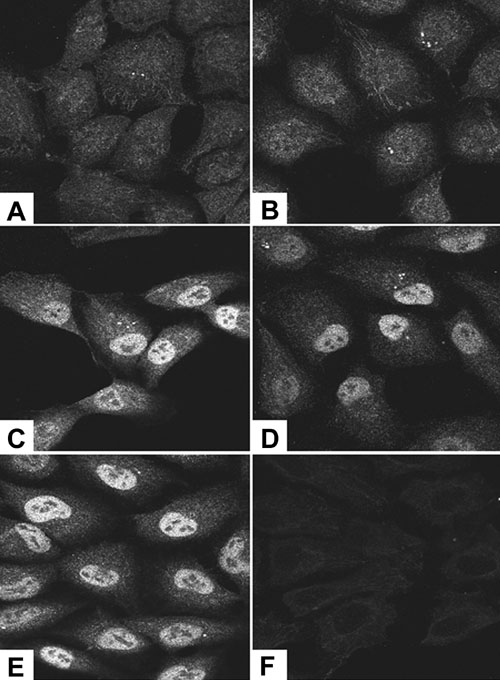
Figure 12. C-fos protein is increased in expression and moves into the nucleus of vitreous-treated retinal pigment epithelial cells
Retinal pigment epithelial (RPE) cells were treated with control medium or vitreous-containing medium for various periods of time. At 30 min, the control (A) and vitreous-treated (B) RPE cells had a similar pattern of staining for c-fos. The pattern of immunostaining remained the same for control cells at 1, 2, and 3 h (data not shown). Vitreous-treated RPE cells at 1, 2, and 3 h (C, D, E) showed intense staining for c-fos in the nucleus. F: Staining with secondary antibody only.