![]() Figure 1 of
Hartung, Mol Vis 2007;
13:66-78.
Figure 1 of
Hartung, Mol Vis 2007;
13:66-78.
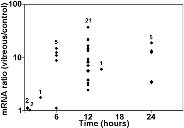
Figure 1. Expression of heme oxygenase-1 mRNA in vitreous-treated retinal pigment epithelial cells
Low passage donor retinal pigment epithelial (RPE) cells were incubated with or without vitreous for up to 24 h. RNA was extracted at each time point and heme oxygenase-1 (HO-1) mRNA was measured by qPCR. Ribosomal protein, large, P0 (RPLP0) was used as an internal standard to correct for small differences in the amount of cDNA used in each reaction. Each point at a particular time indicates a different RPE/vitreous donor pair. The number of donor/vitreous pairs examined at each time point is shown. At 6, 12, and 24 h, REST-XL analysis indicated that HO-1 mRNA was significantly increased (p<0.05) in vitreous-treated RPE cells compared to cells in vitreous-free medium.
