![]() Figure 7 of
Weaver, Mol Vis 2007;
13:707-718.
Figure 7 of
Weaver, Mol Vis 2007;
13:707-718.
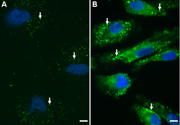
Figure 7. Localization and increased production of ILK in cultured human RPE
Immunocytochemistry with affinity-purified anti-ILK IgG (R3B1) of hRPE plated on glass coverslips. FITC-conjugated donkey anti-rabbit IgG was used as a secondary antibody. A: Non-stressed hRPE stained with R3B1 (green). B: hRPE deprived of serum for 24 h and subsequently stained with R3B1 (green). Increased ILK expression was apparent after stress. Arrows indicate localized ILK staining in hRPE. The scale bar is equal to 1 μm.