![]() Figure 5 of
Weaver, Mol Vis 2007;
13:707-718.
Figure 5 of
Weaver, Mol Vis 2007;
13:707-718.
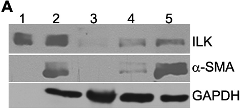
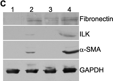
Figure 5. ILK is induced during LEC differentiation and inhibition of ILK retards expression of EMT markers α-SMA and fibronectin
A: Immunoblot of lysates (20 μg/lane) from immortalized and primary mouse LEC that grew out from dissected lens capsules (0-14 days). Lane 1, rhILK (500 ng); lane 2, immortalized LEC; lane 3, primary LEC, 0 days; lane 4, primary LEC, 7 days; lane 5, primary LEC, 14 days. Lysates were probed for ILK and α-SMA as an indicator of EMT. rhILK (lane 1) was used for protein size comparison, and a probe for GAPDH was used to normalize protein loading. B: Densitometry of protein bands from the blot shown in A. Relative expression values are normalized to those of GAPDH. C: Immunoblot of lysates (2 capsules/lane) from primary mouse LEC that grew out from dissected lens capsules (0-14 days). Lane 1, primary LEC + ILK-targeting siRNA, 7 days; lane 2, primary LEC + ILK-targeting siRNA, 14 days; lane 3, primary LEC + GFP-targeting siRNA (control), 7 days; lane 4, primary LEC + GFP-targeting siRNA. Lysates were probed for ILK to verify ILK-targeting siRNA efficiency. Fibronectin and α-SMA were monitored as indicators of EMT. An additional probe for GAPDH was used to normalize protein loading. D: Densitometry of protein bands from the blot shown in C. ILK- and GFP-siRNA-treated explants show a significant difference in protein expression after 14 days of culture (the asterisk indicates a p<0.02). Relative values are normalized to that of GAPDH.