![]() Figure 4 of
Weaver, Mol Vis 2007;
13:707-718.
Figure 4 of
Weaver, Mol Vis 2007;
13:707-718.
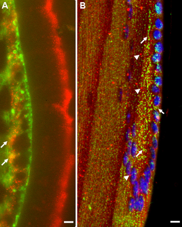
Figure 4. Coincidence of ILK with β1-integrin but not EEA1 in the mouse lens
A: Immunohistochemistry with R3B1 (red) and anti-β1 integrin antibody (green). Yellow indicates coincidence of staining (arrows). The scale bar is equal to 1 μm. B: Immunohistochemistry with R3B1 (green; arrows) and anti-EEA1 antibody (red; arrowheads). No coincidental staining is evident. The scale bar is equal to 10 μm. Morphological alterations and non-specific staining for ILK in the capsule (A, extreme right) occurred with the staining protocol required for integrin immunohistochemistry.