![]() Figure 3 of
Weaver, Mol Vis 2007;
13:707-718.
Figure 3 of
Weaver, Mol Vis 2007;
13:707-718.
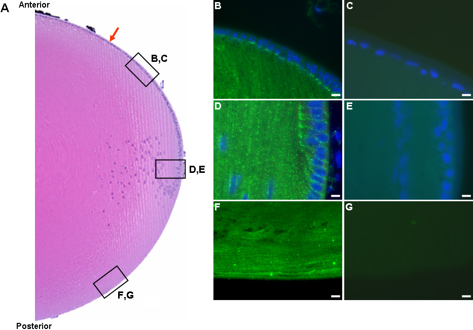
Figure 3. Localization of ILK in the mouse lens
A: Hematoxylin and eosin-stained mouse lens. The red arrow denotes the lens epithelial cell layer. Boxes indicate anterior (B,C), equatorial (D,E), and posterior (F,G) areas imaged for immunohistochemistry. B: Immunohistochemistry of mouse lens anterior LEC and fiber cells with affinity-purified anti-ILK IgG (R3B1). FITC-conjugated donkey anti-rabbit IgG was used as a secondary antibody. C: Immunohistochemistry of anterior LEC with commercial polyclonal anti-ILK antibody. D: Immunohistochemistry of lens equatorial region with R3B1. E: Immunohistochemistry of lens equatorial region with R3B1 pre-absorbed with 10 μg/ml rhILK. F: Immunohistochemistry of the posterior lens with R3B1. G: Immunohistochemistry of the posterior lens with rabbit IgG control. The scale bar is equal to 10 μm.