![]() Figure 2 of
Weaver, Mol Vis 2007;
13:707-718.
Figure 2 of
Weaver, Mol Vis 2007;
13:707-718.
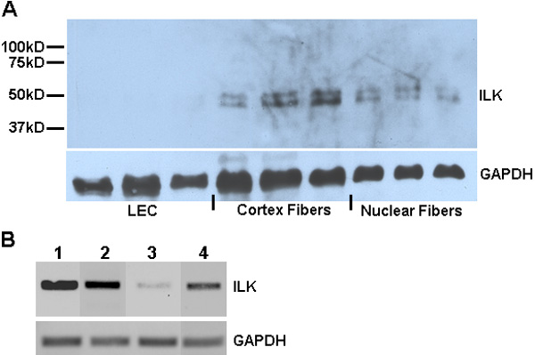
Figure 2. ILK in the mouse lens and in immortalized lens cells
A: Western blot of lysates from cortical and nuclear fiber cells dissected from mouse lenses. Additional probe for GAPDH is shown as a protein loading control. Molecular weight markers are indicated on the left. B: RT-PCR for ILK performed on RNA isolated from immortalized mouse (lane 1) and human LEC (lane 2), as well as mouse lens tissue dissected into capsule/epithelial (lane 3) and lens fiber fractions (lane 4). Probe for GAPDH mRNA is shown as a control for cellular RNA.