![]() Figure 1 of
Weaver, Mol Vis 2007;
13:707-718.
Figure 1 of
Weaver, Mol Vis 2007;
13:707-718.
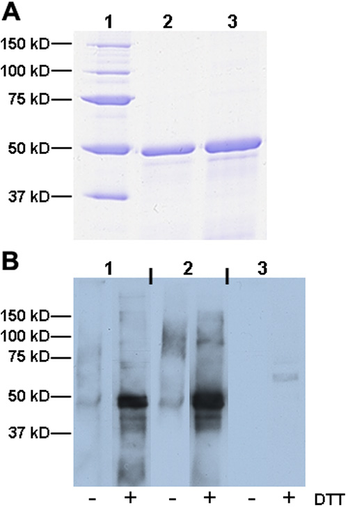
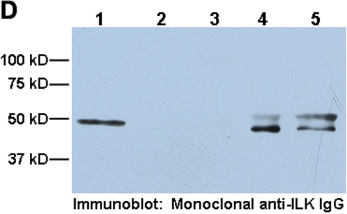
Figure 1. Characterization of polyclonal ILK antibody
A: Purified rhILK used for antigen in rabbits. Lane 1, molecular weight standards; lane 2, 5 μg rhILK; lane 3, 10 μg rhILK. B: Immunoblot of immortalized mouse LEC lysates probed with affinity-purified polyclonal ILK IgG (R3B1; column 1), in comparison with commercially-available anti-ILK monoclonal (column 2) and polyclonal antibodies (column 3), before (-) and after (+) reduction with dithiothreitol (DTT). C: ELISA showing binding of affinity-purified anti-ILK IgG (R3B1) to rhILK (triangle) in comparison with commercially-available anti-ILK polyclonal antibody (circle) and monoclonal antibodies (square). Each point represents three independent experiments, ±SD. D: Comparison of ILK immunoprecipitated from mouse LEC lysates with commercial and R3B1 IgG and subsequently probed with commercial monoclonal antibody. Lane 1, rhILK; lane 2, lysate only (no antibody); lane 3, non-specific rabbit IgG; lane 4, commercial polyclonal antibody; lane 5, R3B1. In A, B, and D, molecular weight markers are shown on the left of each panel.