![]() Figure 8 of
Andrieu-Soler, Mol Vis 2007;
13:692-706.
Figure 8 of
Andrieu-Soler, Mol Vis 2007;
13:692-706.
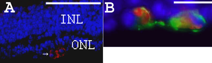
Figure 8. Rhodopsin and β-PDE immunohistochemistry on eye sections from PBS- or WTS ODN-treated rd1 mice at PN28
A: DAPI staining in blue and β-PDE immunostaining in red (arrows) on section from PN28 ODN-treated rd1 retina. B: Combined fluorescence of β-PDE immunostaining in red, rho-4D2 immunostaining in green and DAPI staining in blue on section from PN28 ODN-treated rd1 retina. Scale bars are A, 150 μm; B, 10 μm. β-PDE immunoreactivity is associated with cells in the remaining outer nuclear layer (ONL). This staining appears associated with the cytoplasm, not the nucleus. The rhodopsin immunoreactivity is associated with the same cells in the ONL and the immunoreactivity seems more peripheral to the cytosolic β-PDE staining, suggesting a plasma membrane localization of the rhodopsin immunoreactivity. Relatively few cells in the residual ONL exhibit either rhodopsin or β-PDE immunoreactivity at P28 even after three ODN treatments; however, those remaining positive cells exhibit some degree of clustering.