![]() Figure 7 of
Andrieu-Soler, Mol Vis 2007;
13:692-706.
Figure 7 of
Andrieu-Soler, Mol Vis 2007;
13:692-706.
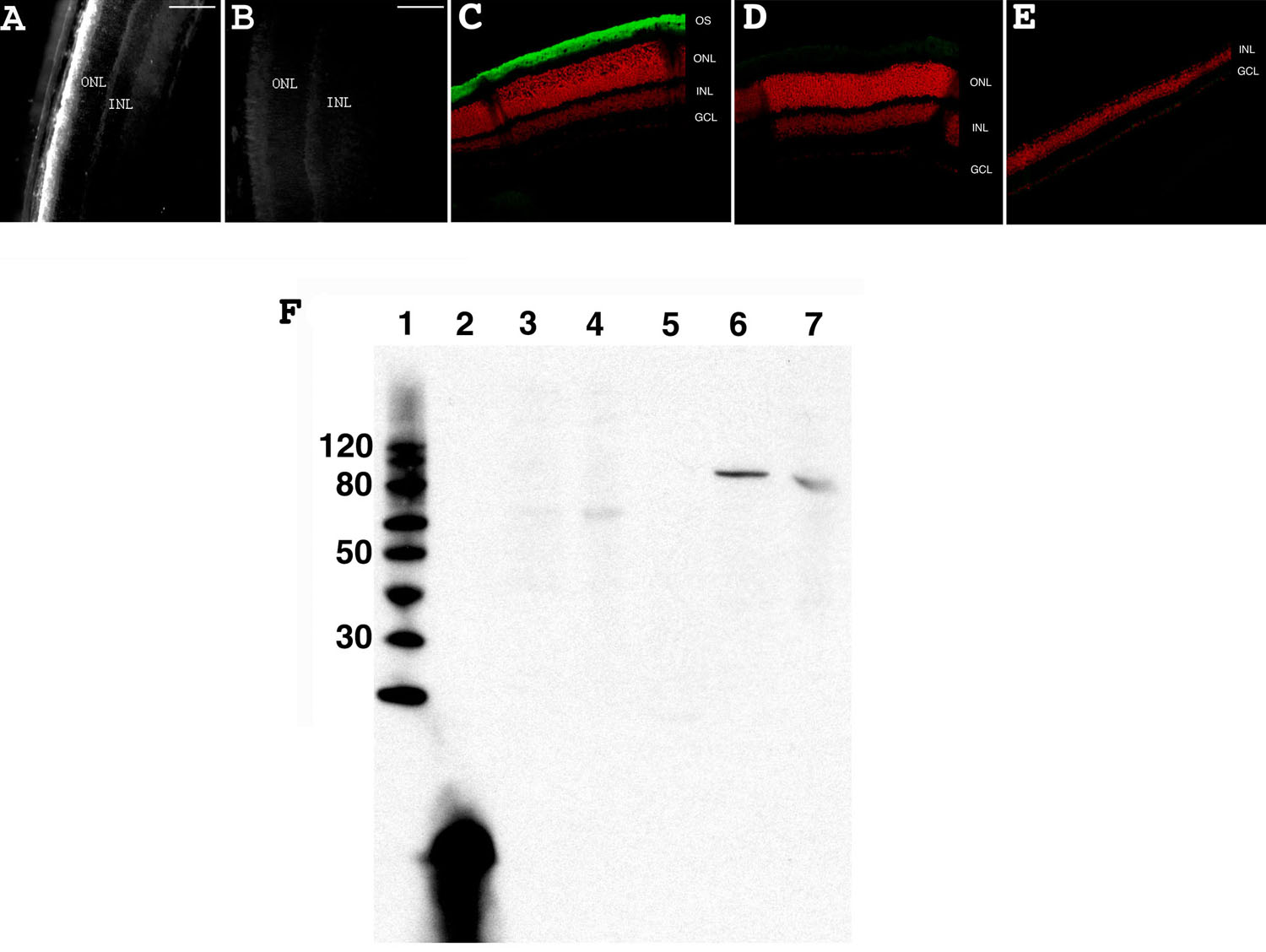
Figure 7. β-PDE immunohistochemistry and western blot
A: Wild type +/+ eye section from a mouse at PN28. B: Control wild type +/+ eye section from a mouse at PN28 using normal rabbit sera. C: Confocal micrograph from a C57/BL6 (wild type; +/+) retina reacted with β-PDE primary antibody and Oregon Green-conjugated goat anti-rabbit IgG (giving green signal in figure) and counter-stained with propidium iodide (giving red signal in figure). D: Confocal micrograph from wild type retina reacted with normal rabbit serum. E: Confocal micrograph from a C3H/henJ mouse (rd1; -/-) retina reacted with β-PDE primary antibody and Oregon green-conjugated goat anti-rabbit IgG. OS indicates outer segments, ONL indicates outer nuclear layer, INL indicates inner nuclear layer, GCL indicates ganglion cell layer. F: Anti-β-PDE "western" immunoblot. Lane 1 is the molecular weight marker (sizes given on left), lane 2 is the polypeptide antigen against which the antibody was raised, lane 3 is protein from a C3H (rd1) mouse retina, lane 4 is protein from a rd10 mouse retina, lane 5 is protein from an FVB (rd1) mouse retina, lane 6 is protein from a CCRC (wild type +/+) mouse retina, lane 7 is protein from a Balb/C (wild type +/+) mouse retina. All mice were at least 30 days old at time of tissue harvest. Scale bars are A, and B, 100 μm. These data suggest that the antibodies are highly specific and selective to β-PDE.