![]() Figure 6 of
Andrieu-Soler, Mol Vis 2007;
13:692-706.
Figure 6 of
Andrieu-Soler, Mol Vis 2007;
13:692-706.
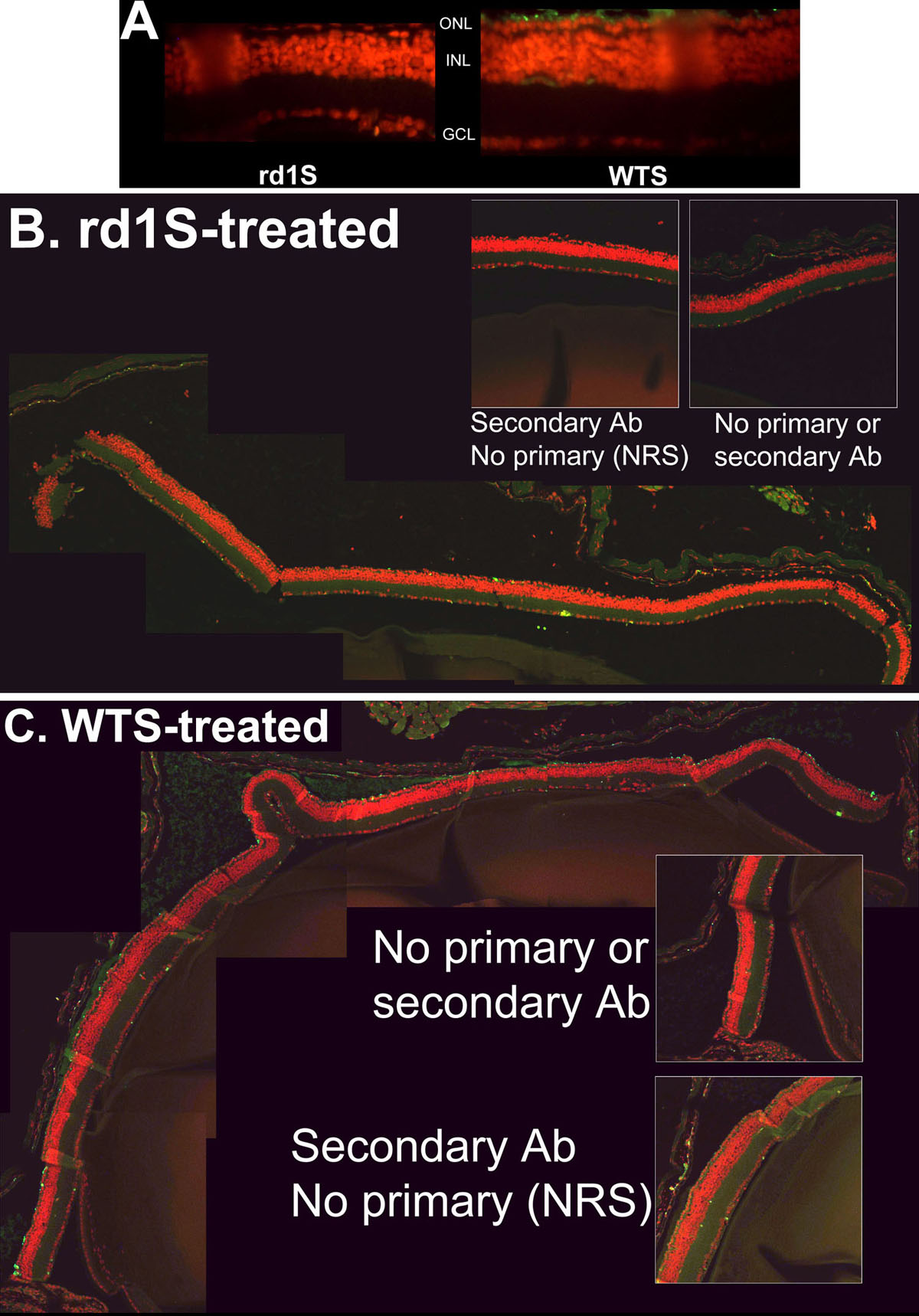
Figure 6. Effect of treatment with WTS versus rd1S at PN33: A confirmatory experiment
In experiments conducted with an independent C3H/henJ colony, littermates treated with WTS ODN had many more rhodopsin-immunopositive cells in the ONL than littermates treated with rd1S, the identical ODN with the exception of having the mutant rd1 nucleotide at the rd1 mutation site. Tissue was harvested and sections prepared at PN33. A: Fluorescent micrographs of retina sections from rd1S-treated mouse (left panel) and WTS-treated mouse (right panel). Rhodopsin immunosignal is green, counterstained nuclei are red. B: Composite image of confocal micrographs of retina section from mouse treated with rd1S. Rhodopsin immunosignal is green, counterstained nuclei are red. Very little rhodopsin signal is apparent. Insets are control sections in which no antibody was used or secondary antibody was used by normal rabbit sera (NRS) was substituted for primary antibody. C: Composite image of confocal micrographs of retina section from mouse treated with WTS. Many rhodopsin-positive cells are apparent in the putative photoreceptor layer. Insets are as in B. Quantification of rhodopsin-positive cells in photoreceptor layers showed that significantly more signal was observed with WTS treatment compared to rd1S treatment (see "Results").