![]() Figure 3 of
Andrieu-Soler, Mol Vis 2007;
13:692-706.
Figure 3 of
Andrieu-Soler, Mol Vis 2007;
13:692-706.
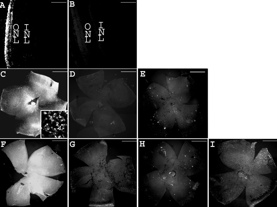
Figure 3. Treatment with WTS preserves rhodopsin at PN28
Rhodopsin immunohistochemistry on wild type eye sections and rd1 whole flat-mount retinas, reflecting the time course of the retinal degeneration and the treatment efficacy. A: Wild type eye section from a mouse at PN28. B: Control eye section from wild type mouse at PN28 using normal mouse serum. C: rd1 flat-mount retina from a mouse at PN19 (inset: high magnification). D: Control flat-mount retina from rd1 mouse at PN19 using normal mouse serum. E: rd1 flat-mount retina from a mouse at PN28. F: PN28 rd1 flat-mount retina injected by WTS with prior iontophoresis at PN4, PN6, and PN8. G: PN28 rd1 flat-mount retina injected by WTS without prior iontophoresis at PN4, PN6, and PN8. H: PN28 rd1 flat-mount retina iontophoresed without oligonucleotide injection at PN4, PN6, and PN8. I: PN28 rd1 flat-mount retina injected with WTSscr7 with prior iontophoresis at PN4, PN6, and PN8. Scale bars are A and B, 100 μm; C, D, E, F, G, H, and I, 1 mm; inset, 10 μm.