![]() Figure 1 of
Andrieu-Soler, Mol Vis 2007;
13:692-706.
Figure 1 of
Andrieu-Soler, Mol Vis 2007;
13:692-706.
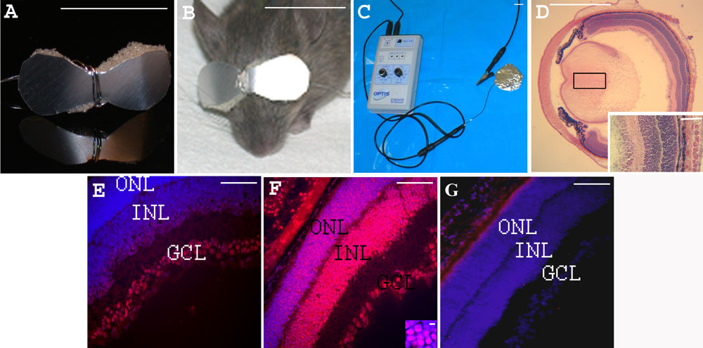
Figure 1. Iontophoresis device and eye sections from PN7 rd1 mice, 1 h after treatment
Iontophoresis device. A: An eye-glass-shaped electrode was made with aluminum foil and single-use disposable medical grade hydrophilic polyurethane sponge. B: The electrode covered both closed eyelids of the treated newborn mouse iontophoresis. C: shows the generator and the return electrode. Eye section 1 h after transpalpebral iontophoresis. D: Hematoxylin and eosin stained eye section showing integrity of the eye structures after iontophoresis Inset shows tissue at high magnification. Eye sections 1 h after intravitreal injection of CY3-tagged oligonucleotide. In E-G, nuclei were stained blue with DAPI and red with CY3. E: without prior iontophoresis, F: with prior iontophoresis (inset: high magnification of the ONL). G: Control retina from an rd1 mouse injected with 1 μl of PBS with prior iontophoresis. The following abbreviations were used: outer nuclear layer (ONL), inner nuclear layer (INL), and ganglion cell layer (GCL). Scale bars: A, B, C, 1 cm; D 1 mm; E, F, G and inset D, 100 μm; inset F, 5 μm.