![]() Figure 4 of
Chandler, Mol Vis 2007;
13:677-691.
Figure 4 of
Chandler, Mol Vis 2007;
13:677-691.
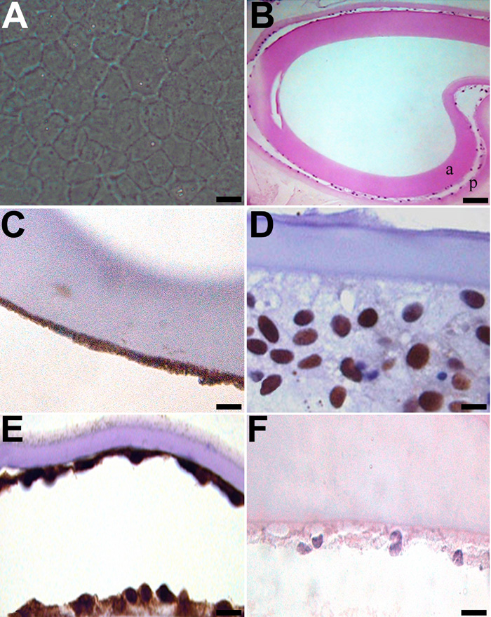
Figure 4. EMT-like changes in the ex vivo lens capsule model
Following sham cataract surgery, canine lens capsules with residual LEC present on the anterior lens capsule were treated with vehicle alone for 2 weeks. By the end of this period, LEC were present on the anterior capsule (a) and had migrated across the posterior lens capsules (p; A and B). These LEC were immunopositive for COX-2, PCNA, and α-SMA (C-E, respectively). To evaluate apoptosis, capsules were stained for cleaved caspase-3 (F). The scale bars are equal to 20 μm, except for B, where the scale bar is equal to 50 μm.