![]() Figure 3 of
Chandler, Mol Vis 2007;
13:677-691.
Figure 3 of
Chandler, Mol Vis 2007;
13:677-691.
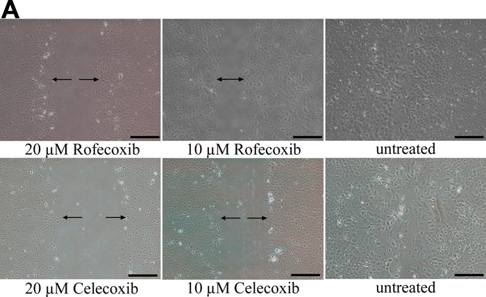
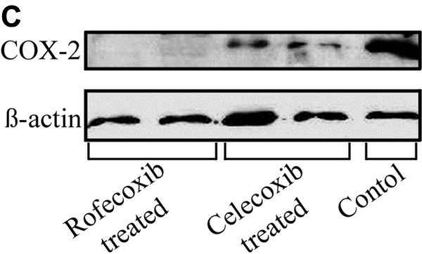
Figure 3. Effects of rofecoxib and celecoxib in migrating LEC in vitro
A: Cells were treated with 10 or 20 μM rofecoxib, celecoxib, or vehicle before a 1 mm scratch was created and LEC were allowed to heal for 24 h. Both rofecoxib and celecoxib treated LEC had decreased rates of wound closure compared to the control. There was a dose dependent response to drug treatment. The scale bar is equal to 0.5 mm. B: There was significantly (p<0.001) decreased healing in LEC cultures treated with either celecoxib or rofecoxib compared to controls. C: Western blot analysis of the LEC treated with 20 μM rofecoxib or celecoxib. There is increased expression of COX-2 in control LEC undergoing EMT-like changes, and decreased expression in LEC treated with the COX-2 inhibitors. There is no expression of COX-2 in rofecoxib treated cells while there is still some COX-2 protein present in the celecoxib treated cells.