![]() Figure 2 of
Chandler, Mol Vis 2007;
13:677-691.
Figure 2 of
Chandler, Mol Vis 2007;
13:677-691.
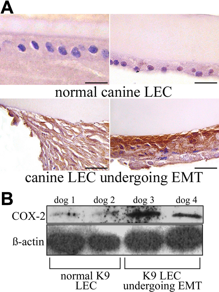
Figure 2. Expression of COX-2 in normal canine LEC and in LEC undergoing EMT-like changes
A: In normal LEC there is little to no COX-2 immunoreactivity, while LECs in cataractous and PCO sample show marked cytoplasmic staining for COX-2, as indicated by diffuse brown cytoplasmic staining. The scale bar is equal to 30 μm. B: Western blot analysis of COX-2 in normal canine LEC and in LEC undergoing EMT-like changes. In normal LEC (dog 1 and 2) there is little COX-2 protein present, while in clinical samples of cataract and PCO (dog 3 and 4) there is increased expression of COX-2 protein. C: Using real-time RT-PCR, expression of COX-2 mRNA was analyzed. In normal LEC there is some expression of COX-2 mRNA, while in clinical samples of cataract and PCO there is a significant (p<0.0145) upregulation of COX-2. Error bars indicate SEM.