![]() Figure 4 of
Joko, Mol Vis 2007;
13:649-658.
Figure 4 of
Joko, Mol Vis 2007;
13:649-658.
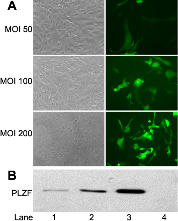
Figure 4. Efficiency of adenovirus vector infection and effect of PLZF on proliferation of cultured HCECs
A: GFP-positive cells in cultured HCECs 48 h after infection. HCECs at 80% confluency were infected at an MOI of 50, 100, and 200 with an adenovirus vector expressing GFP. Phase-contrast photograph (left) and fluorescence photograph (right) showing the infection efficiency was 14% at an MOI of 50, 24% at an MOI of 100, and 41% at an MOI of 200. B: Western blot analysis with anti-PLZF antibody of cultured HCECs infected with Ad-PLZF and uninfected cells. HCECs at 80% confluency were infected with Ad-PLZF. The cells were harvested 48 h after infection. Lane 1: MOI 50; Lane 2: MOI 100; Lane 3: MOI 200; Lane 4: uninfected HCECs. C: The cells were harvested at 24 h after infection, and the cells were seeded into a 16-well strip. Cell index values were determined every hour automatically by the RT-CES system for up to 72 h. Each blot is an average of 8 samples. D: Cell index values at 24 and 48 h are shown. Error bars designate standard deviations. The asterisk indicates a significant difference (p<0.001) between Ad-PLZF and Ad-LacZ (n=8 each).