![]() Figure 3 of
Milla, Mol Vis 2007;
13:639-648.
Figure 3 of
Milla, Mol Vis 2007;
13:639-648.
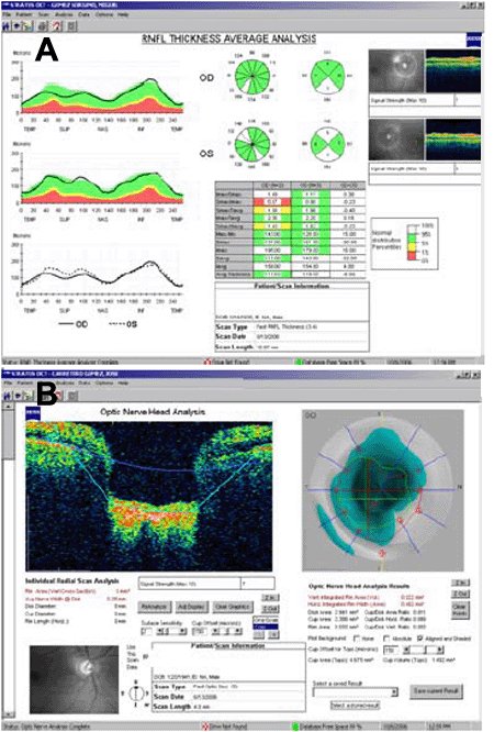
Figure 3. Optical coherence tomography of patients IV-2 and V-1
A: Optical coherence tomography (OCT) diagram for patient IV-2 in which above-normal retinal nerve fiber layer (RNFL) thickness measurements are expressed in white in the pie chart. Green sectors are within the normal range. B: OCT Fast optic nerve head protocol of patient V-1, confirming cup enlargement.