![]() Figure 5 of
Spence, Mol Vis 2007;
13:57-65.
Figure 5 of
Spence, Mol Vis 2007;
13:57-65.
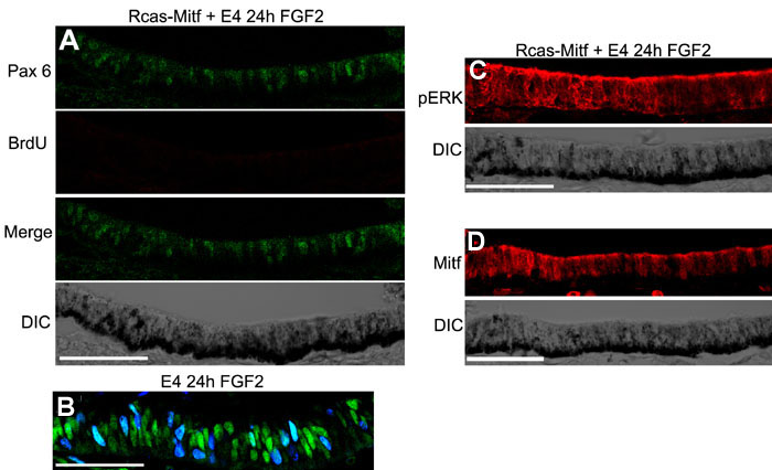
Figure 5. Mitf inhibits transdifferentiation by inhibiting Pax6, but not FGF signaling
A: Expression of Pax6 (top), BrdU (second), Pax6 and BrdU merge (third), and the DIC image (bottom) of the RPE after subretinally injecting Rcas-Mitf at E3, removing the retina at E4 and exposing the RPE to FGF2 for 24 h. B: Expression of Pax6 and BrdU in the RPE after 24 h of exposure to FGF2 and retina removal at E4. C: Erk phosphorylation in the RPE after subretinally injecting Rcas-Mitf at E3, removing the retina at E4 and exposing the RPE to FGF2 for 24 h (top), and the corresponding DIC image of the RPE (bottom). D: Mitf protein levels in the RPE after subretinally injecting Rcas-Mitf at E3, removing the retina at E4 and exposing the RPE to FGF2 for 24 h (top), and the corresponding DIC image of the RPE (bottom).