![]() Figure 1 of
Chiang, Mol Vis 2007;
13:635-638.
Figure 1 of
Chiang, Mol Vis 2007;
13:635-638.
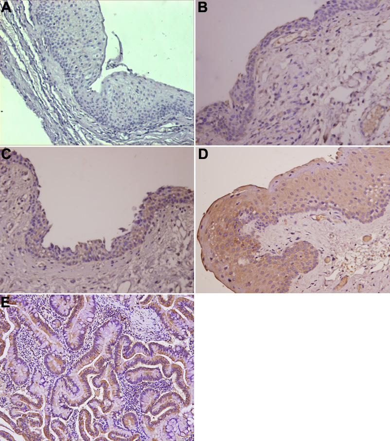
Figure 1. Immunohistochemical analysis of COX 2 protein expression in pterygium
A: This panel shows negative expression. Positive immunostaining of scores +, ++, and +++ were showen in B, C, and D. E: COX 2 protein expression in colon cancer tissue was used as a positive control. COX 2 immunostaining showed a brown reaction product, and COX 2 immunoreactive protein was seen predominantly over the basal epithelial layer. No substantial staining was visible in the subepithelial fibrovascular layers. COX 2 staining was evident in cytoplasm and membrane of the epithelial layer.